Nasolabial folds: what forms them, what deepens them, what moves them
·21 min read·Updated
The first thing to fix about nasolabial folds is the category. They are filed everywhere as wrinkles, treated as damage, and priced as something to remove. But a nasolabial fold is not a crease worn into skin by use. It is a structural boundary that was there before anything happened to your face — visible in infants, visible in every healthy twenty-year-old, and visible in the people selling its removal. What changes with age is not whether you have one. It is how deep it sits when your face is doing nothing.
That distinction decides everything else, because it sorts the interventions. Things that act on skin surface are aimed at the wrong tissue. Things that act on volume above the fold are aimed at the right one. And the exercises are aimed at a muscle that, if it did what the article promised, would make the fold more prominent rather than less.
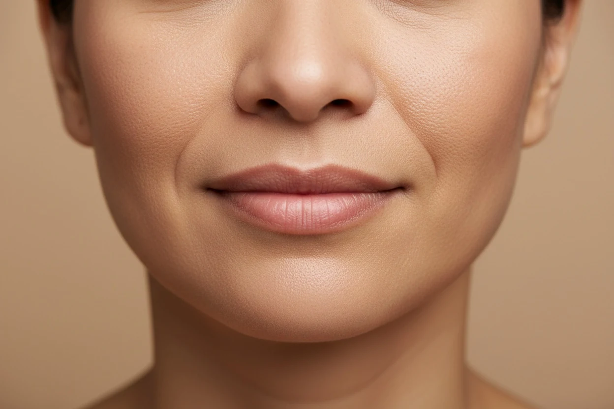
What nasolabial folds are, and how they differ from smile lines
Barton and Gyimesi dissected the region and published the anatomy in Plastic and Reconstructive Surgery in 1997. The fold marks the line where muscles of the upper lip — the levator group and zygomaticus — insert into the dermis. Skin that is anchored to muscle sits at a different level from skin that is not, and the resulting step is the fold. Cheek tissue sits on the outer side of that step and lip tissue on the inner side, so the boundary is the seam between two different mechanical territories.
This is why the terminology in the SERP results is a mess and worth straightening out, because the three words are used for three different things.
| Term | What it names | Present in a young face? |
|---|---|---|
| Nasolabial fold | The tissue boundary from the side of the nose to the mouth corner — a step in the surface | Yes, always |
| Nasolabial crease or line | The etched line that can develop within that boundary over time | Usually not |
| Smile or laugh lines | Everyday usage, applied loosely to both, and sometimes to the radial lines around the eyes instead | Depends who is speaking |
| Marionette lines | A different structure entirely — from the mouth corner down to the jawline, below the fold | Rarely |
What causes nasolabial folds to deepen
The modern account of midface ageing is compartmental. Rohrich and Pessa showed in 2007 that facial fat is not one continuous layer but a set of discrete compartments separated by septa, each with its own blood supply — which immediately explains why a face ages unevenly rather than deflating as a whole.
Gierloff and colleagues then measured what happens to those compartments over time. Their 2012 study took computed tomography of cadaver heads in two age bands and quantified the change. Three findings matter here, and all three point at the fold.
- The compartments migrate downward. The distance between the fat compartments and the infraorbital rim was greater in the older group — the midface volume physically sits lower.
- Volume redistributes inside each compartment. The lower third of each compartment grew in sagittal diameter while the upper third shrank, so even without net loss, mass accumulates at the bottom edge.
- Specific compartments lose volume, notably the deep medial cheek fat, which is precisely the support that sits behind the upper portion of the fold.
Put those together and the mechanism is not mysterious. Tissue above the fold sags towards a boundary that is anchored and cannot move with it, and mass piles up on the cheek side of a fixed seam. The step gets taller. Nothing was worn into the skin.
The bone underneath moves too
Mendelson and Wong reviewed skeletal ageing in 2012 and described what happens to the facial skeleton as a scaffold: the maxilla loses projection, the pyriform aperture around the nose widens, and the orbital rim recedes. Soft tissue that was draped over a well-projected structure ends up draped over a less projected one, with the same surface area and less to hold it forward. Fold depth is the visible consequence of a change several millimetres beneath it.
Descent or deflation — the argument that changes the answer
This is where the field is genuinely divided, and it is worth understanding because it determines which interventions make sense. The traditional account is descent: tissue falls, and lifting it is the fix. Lambros challenged that in 2007 with a study comparing photographs of the same individuals decades apart and found that landmarks he expected to have descended had largely stayed put — what changed was volume, not position. Ageing as deflation rather than as gravity.
The current position takes both. Gierloff's imaging supports real inferior migration; Lambros's photographs support real deflation; most faces are showing some of each. It has a practical consequence: a fold driven mainly by lost volume and a fold driven mainly by descended tissue respond to different things, and no article can tell you from a distance which yours is. A clinician looking at your face at rest and in animation can.
How nasolabial fold severity is graded
Trials in this area report their results on the Wrinkle Severity Rating Scale, validated by Day and colleagues in 2004. It is worth knowing because it converts a subjective worry into a category, and because every efficacy number quoted anywhere on the internet is a movement along it.
| Grade | Description | What it looks like |
|---|---|---|
| 1 — Absent | No visible fold, continuous skin line | Uncommon in adults at any age |
| 2 — Mild | Shallow but visible flattening with a subtle indentation | The typical young adult fold |
| 3 — Moderate | Moderately deep fold, clear facial feature, less prominent when stretched | The most common reason people search this |
| 4 — Severe | Very long and deep fold, prominent feature, under 2 mm of visible fold when stretched | Where clinical intervention is usually discussed |
| 5 — Extreme | Extremely deep and long, detrimental to facial appearance, 2–4 mm of visible V-shaped fold when stretched | Rare outside significant volume loss |
Do smile lines come from smiling?
The most repeated piece of folk advice in this area — smile less, it is carving lines into your face — has the causation reversed. Smiling contracts exactly the muscles that insert along the fold, so it makes the boundary visible. That is a demonstration of where the anchor points are, not a mechanism for deepening them. If expression were the driver, the deepest folds would belong to the most expressive people, and nobody has shown that.
There is one nuance worth keeping. Repeated contraction does contribute to etched surface lines within a fold over decades, the way it does around the eyes. That is the crease from the terminology table, not the fold, and it is the component that surface treatments can reach. The structure remains a structure.
A face is not damaged by the expressions it makes. The fold marks where your muscles attach, and it was drawn before you had anything to smile about.
Nasolabial folds in your 20s: why they show up early
A substantial share of the people searching this are nowhere near an age where compartment migration explains anything, and the standard ageing article does not answer them. A visible fold at 22 is not early ageing. In a young face the fold is usually about where things attach and how much sits above them.
- Insertion pattern. Where your levator muscles meet the dermis is individual anatomy. A more superficial insertion gives a more defined boundary in a face with no ageing changes at all.
- Full midface volume. More cheek tissue folding over a fixed seam produces a deeper step. This is the case where the fold is a consequence of fullness rather than of loss, and where volume-adding approaches are pointed the wrong way.
- Maxillary projection. A shallower midface skeleton gives less forward support, which shows up decades before any bone resorption. This is skeletal, and it is also why the shape of the lower and middle thirds of your face is worth measuring before assuming a fold is an ageing sign.
- Expressiveness plus thin skin. Produces a crisp line early. This is the profile where the surface-quality interventions — sunscreen, retinoids — are best matched to the actual finding.
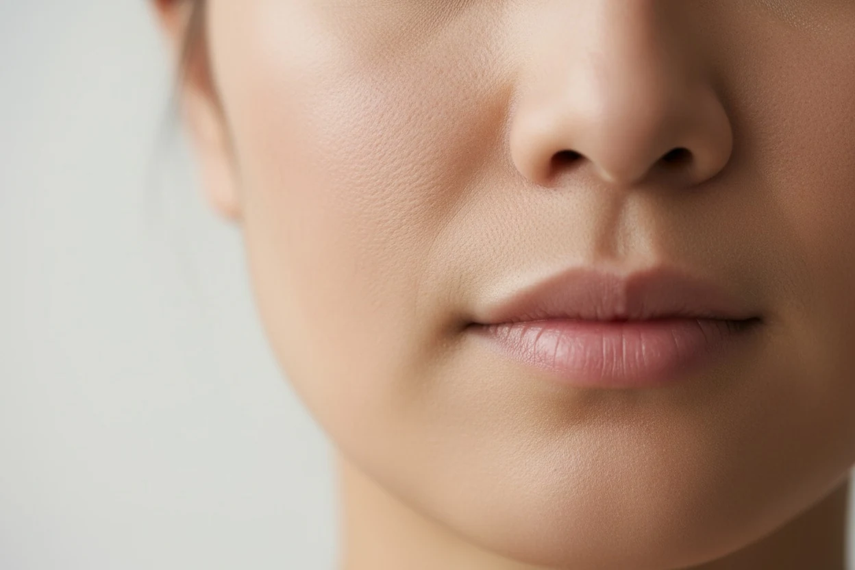
The at-rest test separates these from ageing cleanly. Relax your face completely, no smile, mouth closed, and photograph straight on. A young structural fold typically softens almost to nothing at rest; an age-related one persists because the volume above it has moved and stays moved.
| Driver | What you see at rest | What acts on that driver |
|---|---|---|
| Muscle insertion pattern | A defined boundary that softens when the face relaxes | Nothing needs to — it is how your face is built |
| Full midface | A deeper step on smiling, soft at rest | Not added volume, which adds to the cause |
| Shallow maxilla | A fold present since the teens, not getting deeper | Nothing topical; it is skeletal |
| Thin skin with expressive muscles | A crisp line that fades at rest | Sunscreen and retinoids, on the skin component |
| Fat compartments moving down (with age) | A fold that stays when the face is relaxed | Volume support, as a clinical decision |
| Maxillary bone losing projection (with age) | A fold that deepens slowly over decades | Volume support, as a clinical decision |
What age is it normal to get nasolabial folds?
Every age. The fold is present in infants, so the useful question is not when it appears but how deep it is at rest. The mild grade on the severity scale above — a shallow but visible indentation — describes the ordinary young-adult fold. We did not find a study giving a reliable age-by-age distribution of grades, so no page, including this one, can honestly tell you your fold is early for your age.
Should you treat a fold in your 20s?
Nothing in the research cited here says you need to. The filler studies in this guide enrolled adults with established folds, and none of them tested filler as a way of preventing a young fold from deepening later — so claims that early filler "stops the cascade" are marketing rather than findings. Where the driver is full cheeks, adding volume is pointed the wrong way. The two things with good evidence at this age are the preventive ones further down: daily sunscreen and not smoking.
Nasolabial fold treatments, ranked by what was measured
This is the section that most pages get wrong by listing things in order of popularity. Ordered by evidence quality instead, the picture changes considerably — and two of the strongest entries are preventive rather than corrective.
| Intervention | Tissue it acts on | Best available evidence |
|---|---|---|
| Hyaluronic acid filler | Volume, injected beneath the fold | Strongest. 2022 meta-analysis, 14 randomised trials, 1,190 patients, graded on WSRS |
| Daily broad-spectrum sunscreen | Skin quality, preventively | Strong, and randomised. 4.5-year community trial, 903 adults, 24% less photoageing |
| Topical retinol | Dermal collagen and glycosaminoglycans | Good. Randomised, double-blind, vehicle-controlled; measured on fine wrinkling, not fold depth |
| Stopping smoking | Skin and midface soft tissue | Good, observational. 79 identical twin pairs; smoking twins scored worse specifically on nasolabial folds |
| Calcium hydroxylapatite placed over the cheekbone | Volume above the fold, not in it | Limited. Retrospective study, 51 women; fold length shortened by 13.5% on one side and 6.2% on the other at six months |
| Other injectables (poly-L-lactic acid) | Volume, gradually | Trials exist, fewer head-to-head comparisons than for HA |
| Botulinum toxin injected into the skin | Fine lines and texture, not volume | Small. Split-face randomised trial, 18 people; fold wrinkles improved for 12 weeks. Industry-funded |
| Surgical lifting and fat grafting | Position and volume together | Established clinically; the intervention with the largest effect and the largest risk |
| Thread lifting | Position | An active research area; longevity and comparative data are still thin |
| Facial roller or gua sha | Skin elasticity, muscle tone | One 2025 randomised trial, 34 women — measured contour, never fold depth |
| Facial exercise | Muscle | No controlled evidence for this fold. 2014 review found no randomised trials at all |
| Creams claiming to erase folds | Skin surface | None. A cream cannot reach a structural boundary or add midface volume |
What the filler meta-analysis actually found
Peng and colleagues pooled 14 randomised controlled trials covering 1,190 patients in 2022, and the question they asked was narrower and more useful than 'does filler work'. They compared monophasic against biphasic hyaluronic acid on WSRS improvement, satisfaction and adverse events. Monophasic came out ahead on fold improvement and on the proportion of satisfied patients, with no significant difference in adverse event rate between the two.
That is a product-selection finding, and it is the level of detail worth carrying into a consultation rather than out of one. This region also sits near the facial artery and its branches, which is why injection here carries a documented vascular risk profile and why the decision is a clinical one. Nothing on this page is a recommendation to have it, and no page can tell you whether it is appropriate for your face.
The two preventive entries deserve their ranking
Hughes and colleagues randomised 903 adults in Queensland to daily or discretionary sunscreen for four and a half years, with blinded assessment of skin microtopography. The daily group showed no detectable increase in skin ageing over the whole period, and 24% less than the discretionary group. It is one of very few randomised trials of anything in cosmetic dermatology, and the intervention costs almost nothing.
Kafi and colleagues ran a randomised, double-blind, vehicle-controlled trial of 0.4% retinol applied three times weekly for 24 weeks, with each participant's other arm as the control. Fine wrinkling improved significantly against vehicle, and biopsies showed increased glycosaminoglycan expression and procollagen I. The endpoint was fine wrinkling on aged skin rather than nasolabial fold depth — so it supports the skin-quality half of the picture, which is the etched line, not the structural step.
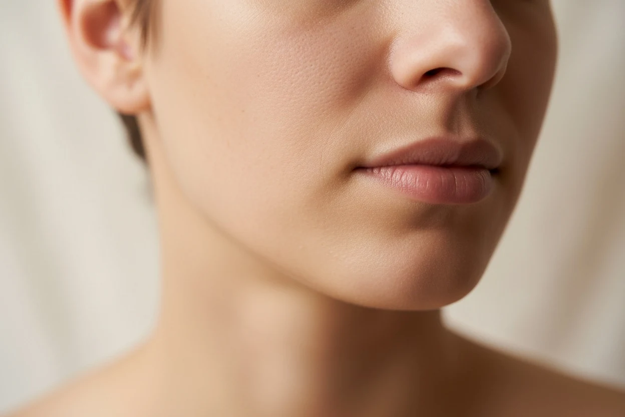
Skin quality matters more here than people expect. Fink, Grammer and Matts found that changing skin colour distribution alone shifted ratings of age, health and attractiveness with no change to the underlying geometry at all. A softer-reading fold is partly a lighting-and-surface phenomenon, and surface is the part you can act on cheaply.
Nasolabial fold filler: what trials measured, how long it lasted, and the risk
Filler is the most studied intervention for this fold, which is exactly why it deserves a section that puts the result, the duration and the risk side by side. Nothing here is a recommendation to have it; it is what was measured, so the questions you ask in a consultation can be better ones.
Filling the fold vs supporting the cheek above it
There are two different targets, and the anatomy at the top of this guide explains why. Filling the fold itself puts volume into the step. Supporting the midface above it puts volume where the fat compartments have moved from, so less tissue sits on the cheek side of the seam. A 2025 retrospective study of 51 women aged 30 to 55 who had calcium hydroxylapatite placed over the cheekbone area only, not in the fold, measured the fold six months later: its length shortened by about 13.5% on one side and 6.2% on the other, with severity scores improving too. It had no control group, so it shows that the indirect route moves the fold, not how it compares with filling it directly.
How long nasolabial fold filler lasted in trials
Durations quoted online are usually a range with no source. One of the better-designed trials gives a concrete answer for one hyaluronic acid product: 87 people with severe folds had it injected on one side and bovine collagen on the other, and the folds were graded on a five-point scale every four weeks. At 24 weeks, 96% of the hyaluronic acid-treated folds kept a clinically significant correction, and 81% kept it for a year or more. Repeat treatment needed less than half the volume of the first. Two caveats belong next to those numbers: the trial was run with the manufacturer, and results for one product in severe folds do not transfer automatically to other products or milder folds.
The risks that belong in the same paragraph
The nasolabial region sits over branches of the facial artery, and filler that enters or compresses a vessel can block blood flow. The rarest and most serious outcome is vision loss. A review of every published case up to 2015 found 98 cases of vision changes after filler, and the nasolabial fold was the injection site in 13.3% of them; an update covering 2015 to 2018 found 48 more, 14.6% from the nasolabial fold, with hyaluronic acid responsible for 81% of the newer cases. In both reviews, no treatment reliably restored sight, and most people did not fully recover their vision. The absolute risk is small — these are case reports out of a very large number of injections — but it is the reason this region is a medical procedure and not a beauty treatment.
Hyaluronic acid has one safety property the other fillers lack: an enzyme, hyaluronidase, can dissolve it. Current guidance treats vascular occlusion as an emergency needing immediate high-dose hyaluronidase, and uses lower doses for non-urgent problems such as lumps. Whoever injects should have it on hand and know how to use it, which is a fair thing to ask before anything is injected.
Botox or filler for nasolabial folds?
They act on different tissue, so the question is less which is better than which problem each can reach. Filler adds volume. Botulinum toxin relaxes muscle, or, injected into the skin rather than the muscle, acts on the skin itself — it adds nothing. Since the fold deepens mainly through volume moving, toxin is not a routine treatment for the fold itself.
| Botulinum toxin | Hyaluronic acid filler | |
|---|---|---|
| Acts on | Muscle activity; injected into the skin, the skin | Volume under or above the fold |
| What a trial measured | Into the cheek skin: fine lines of the fold improved for 12 weeks in an 18-person split-face trial | Fold severity on a five-point scale; correction held a year or more in 81% of severe folds in one trial |
| What it cannot do | Replace the volume whose shift deepens the fold | Remove the boundary itself |
| Main risk here | Weakening the muscles that lift the lip and shape the smile; one participant developed facial palsy at the higher dose | Vascular occlusion; the nasolabial fold is among the reported sites of filler-related vision loss |
How to get rid of nasolabial folds without fillers
The honest first sentence: you cannot remove a structure you were born with, with or without filler. What non-injectable options can change is the etched crease within the fold and the skin over it, and — preventively — how fast the fold deepens. In order of evidence, that means daily sunscreen, not smoking, and a retinoid, with everything else a long way behind.
Face yoga for nasolabial folds: the muscle-volume claim
The usual claim is that exercising the cheek muscles builds them up so they fill out the fold. There are two problems with it. No controlled trial has measured fold depth after facial exercise; the systematic review found no randomised trials at all, and the best pilot study rated cheek fullness, not the fold. And the fold forms where muscles of the upper lip pull on the skin, so more muscle bulk on the cheek side of that seam is not obviously the direction you want. The facial exercise guide covers the exercise evidence in more detail.
Thread lifts, and why they come up in Korean clinics
Threads placed under the skin to reposition tissue are a common answer to the question of what Koreans do for nasolabial folds, and some of the most detailed recent writing on them comes from Korean anatomists. A 2024 review from Yonsei University classifies nasolabial folds into three types by cause and describes thread techniques for each, with attention to the arteries that make the area risky. It is a technique guide rather than an outcome trial, which matches the treatment table above: threads are an active area, and long-term comparative data are still thin.
Creams and serums: retinoids, vitamin C, peptides
Retinoids are the only topical group with solid evidence of changing the skin itself. In the classic tretinoin study, photodamaged skin made 56% less new collagen than sun-protected skin, and ten to twelve months of prescription tretinoin raised collagen formation by 80% while the placebo cream group fell by 14%. The retinol trial earlier in this guide found the same direction with an over-the-counter strength. Both measured arm skin, not the fold, so they support softening the crease component rather than the step. We did not find a controlled trial showing vitamin C serums or peptide creams changing nasolabial fold depth, and a cream cannot reach the volume beneath the fold.
How to prevent nasolabial folds from deepening
Four, in the order the evidence supports rather than the order they are usually listed.
- 1Daily broad-spectrum sunscreen. The randomised trial above is the strongest result in this entire subject, and it is preventive — it does nothing for a fold that already exists, and a great deal for the one you would otherwise have in ten years.
- 2Not smoking. Okada and colleagues photographed 79 pairs of identical twins where one smoked or had smoked at least five years longer, and had three blinded judges grade them. The smoking twins scored worse on nasolabial folds specifically, alongside lower lid bags, malar bags, upper lip wrinkles and jowls, with effects concentrated in the middle and lower thirds of the face. Genetics held constant, five years of difference was visible.
- 3Weight stability. Repeated substantial gain and loss stretches and empties the same compartments Gierloff measured. There is no trial isolating this against fold depth, so it sits below the two above on evidence and above them on how often it is ignored.
- 4Sleep position. Anson, Kane and Lambros described the mechanism in 2016 — hours of nightly compression and shear against a pillow — and the twin study found prone sleepers had measurably more facial asymmetry. Neither measured this fold as an endpoint. If you are wondering how to get rid of nasolabial folds while sleeping, sleeping on your back removes one source of compression — a variable, not a treatment. Worth changing if you can; not the lever people hope it is.
Why nasolabial folds look deeper in photos
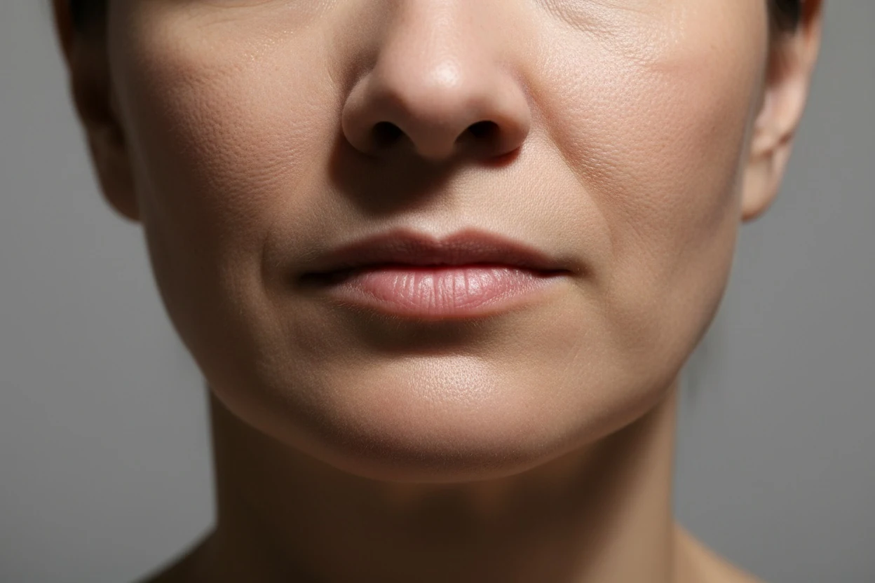
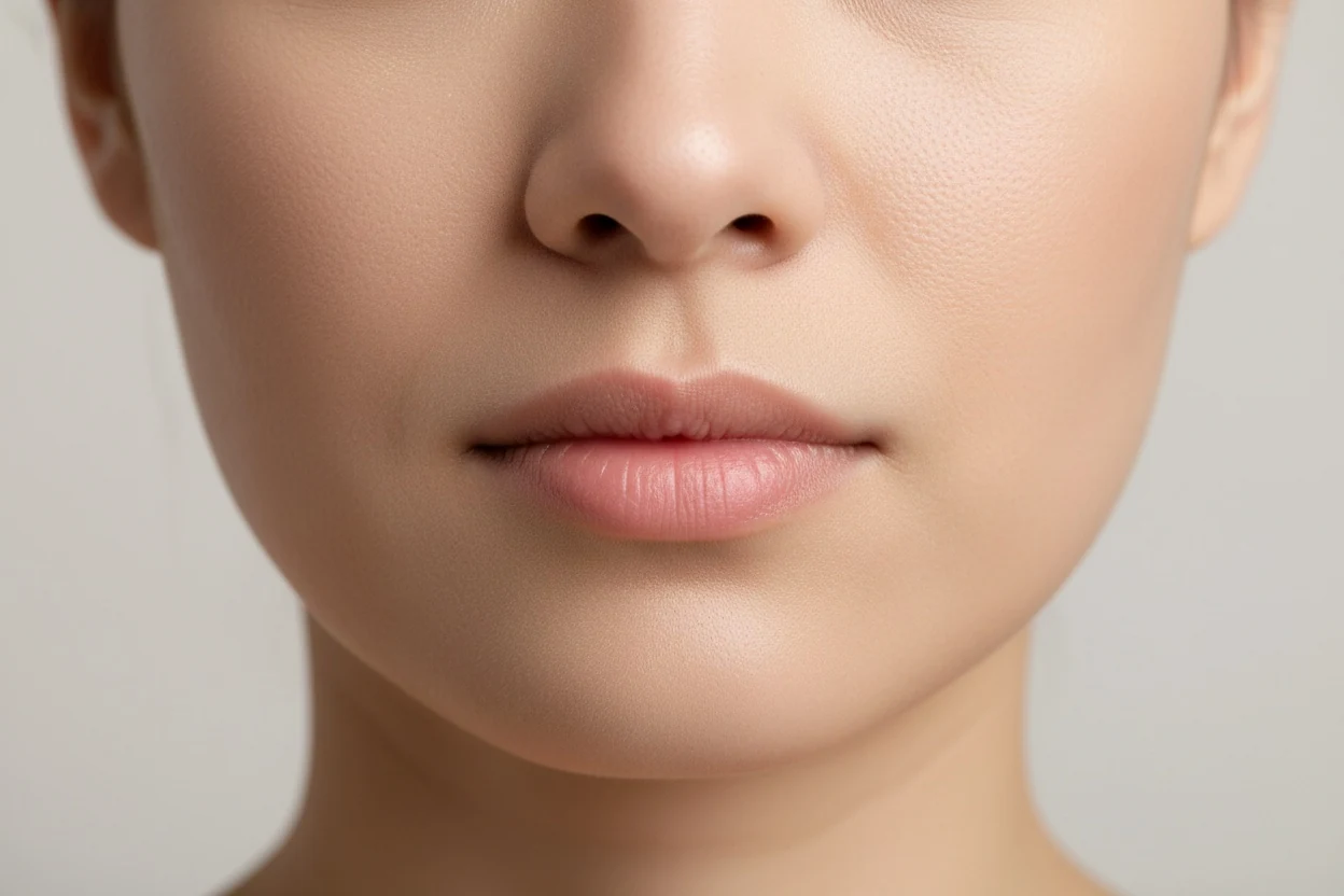
More people are alarmed by a photograph than by a mirror, and the reason is usually the light. This region is read almost entirely as a shadow, which means the light source has more control over its apparent depth than the tissue does.
| Condition | Effect on the apparent fold | Why |
|---|---|---|
| Overhead lighting | Substantially deeper | The shadow falls directly into the boundary — this is the single biggest artefact |
| Hard side light | Deeper on the shadowed side, shallower on the lit side | Manufactures an asymmetry that is not in the face |
| Flat, even frontal light | Shallowest and most honest | Minimal shadow, so what remains is structure |
| Camera held low | Deeper | Foreshortens the midface and stacks tissue towards the fold |
| Short lens held close | Deeper | Perspective distortion pushes the midface forward relative to the mouth |
| Smiling | Much deeper | Muscle contraction pulls directly on the insertion line |
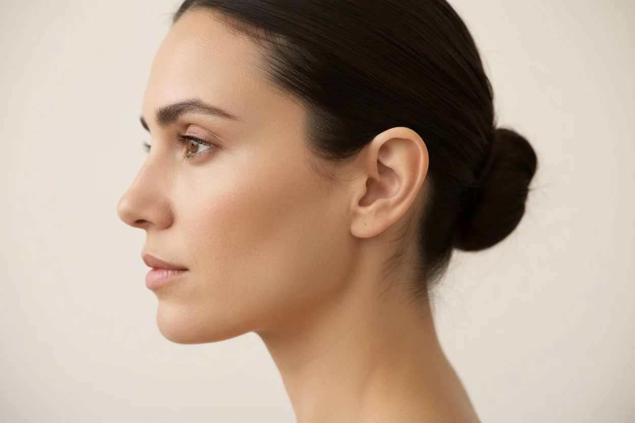
There is one genuine limit on top of that. This fold is partly a depth feature, and a single front-on photograph contains no depth information at all — the projection of the maxilla behind it is invisible head-on. It is the same limitation that means four of the seven nose types cannot be settled from a front photo either. A three-quarter view carries more of what matters here than a straight-on one does.
The age test reports the proportions that actually move with time — the lower third against the other two, the philtrum, and where your measurements sit relative to the ranges for your age band. It runs in your browser and your photo is never uploaded.
See which proportions have shiftedSeeing a nasolabial fold change before deciding on one
If you are far enough along to be weighing an actual procedure, the useful thing is not another article. It is seeing the change on your own face rather than on a stock before-and-after belonging to someone with different anatomy.
The midface volume simulation renders one change on your own photograph so you can look at the result rather than imagine it. Two things about that render are worth stating plainly, because they are the reason it is useful at all: it is an AI simulation and not a surgical or clinical outcome, and every frame on this site says so. It is something to take into a consultation as a way of describing what you want, not a substitute for having one, and not a prediction of what any clinician would achieve.
And the last thing, which the anatomy has been saying throughout: this page is a description of published research, not medical advice, and no measurement or image on this site is a diagnosis. Whether anything here applies to your face is a question for a qualified clinician who is looking at it.
Frequently asked questions
What causes nasolabial folds?
The fold itself is anatomy: a boundary where muscles of the upper lip insert into the skin, present from birth and visible in babies. What deepens it with age is a volume shift above and behind it. Computed tomography of the midface shows the fat compartments migrating downward and redistributing within themselves, while the maxillary bone underneath loses projection. Skin thinning and sun damage then make the resulting shadow crisper. Smiling reveals the fold; it does not build it.
Can you get rid of nasolabial folds completely?
No, and any method promising it is promising to remove a structure that healthy young faces also have. The realistic question is how deep the fold is at rest and how sharply it reads, both of which are gradeable on a validated five-point scale. Softening a grade or two is what the published trials actually measure; erasure is not an endpoint anyone reports.
Why do I have smile lines at 20?
Almost always because of where your muscles insert and how much midface volume you carry, neither of which is age-related. A shallow maxilla, full cheeks that fold over a fixed boundary, thin skin, or simply expressive muscles will all produce a visible fold in a face with no ageing changes at all. The distinguishing test is whether it is there when your face is completely at rest — a young fold usually softens almost to nothing, while an age-related one persists.
Do facial exercises work on nasolabial folds?
There is no controlled trial showing that they do. The 2014 systematic review of facial exercise for rejuvenation found nine reports, none randomised or controlled, and concluded the evidence was insufficient. The 2018 Northwestern pilot did report improved cheek fullness ratings after twenty weeks of daily work, which is the closest anything comes, but it rated fullness rather than fold depth. Exercise increasing the volume of a muscle that sits above a fold is also not obviously the direction you want.
Which filler is used for this area and what does the evidence say?
Hyaluronic acid gel is the most studied. A 2022 meta-analysis pooled 14 randomised trials and 1,190 patients, comparing monophasic against biphasic formulations on the Wrinkle Severity Rating Scale, and found monophasic performed better on both fold improvement and patient satisfaction with no difference in adverse events. That is a comparison between products, not a recommendation to have one — injection near this region carries vascular risk and the decision belongs with a qualified clinician.
Does Botox work on nasolabial folds?
Not on the fold's cause. Toxin relaxes muscle or, injected into the skin, acts on the skin; it adds no volume, and the fold deepens mainly because volume has moved. A small split-face trial of toxin injected into the cheek skin found the fine lines of the fold improved for 12 weeks, and one participant developed facial palsy at the higher dose. It is a narrow tool with a smile-changing risk, and a decision for a clinician.
How long does nasolabial fold filler last?
It depends on the product and the fold. In one randomised within-subject trial of a hyaluronic acid filler in severe folds, 96% of treated folds held a clinically significant correction at 24 weeks and 81% held it for a year or more. That trial was run with the manufacturer, and other products and milder folds may behave differently.
Does sleeping on your side make the crease worse?
It is a plausible contributor with supporting observational work rather than a proven cause. Research on sleep distortion describes the face being compressed and shear-loaded for hours nightly, and a twin study found prone sleepers had measurably more facial asymmetry. Neither study measured fold depth as its endpoint, so the honest summary is that sleep position is on the list of things worth changing and not at the top of it.
References
- Barton, F. E. Jr., & Gyimesi, I. M. (1997). Anatomy of the nasolabial fold. Plastic and Reconstructive Surgery, 100(5), 1276–1280. PMID 9326792.
- Rohrich, R. J., & Pessa, J. E. (2007). The fat compartments of the face: anatomy and clinical implications for cosmetic surgery. Plastic and Reconstructive Surgery, 119(7), 2219–2227. PMID 17519724.
- Gierloff, M., Stöhring, C., Buder, T., Gassling, V., Açil, Y., & Wiltfang, J. (2012). Aging changes of the midfacial fat compartments: a computed tomographic study. Plastic and Reconstructive Surgery, 129(1), 263–273. PMID 21915077.
- Mendelson, B., & Wong, C. H. (2012). Changes in the facial skeleton with aging: implications and clinical applications in facial rejuvenation. Aesthetic Plastic Surgery, 36(4), 753–760. PMID 22580543.
- Lambros, V. (2007). Observations on periorbital and midface aging. Plastic and Reconstructive Surgery, 120(5), 1367–1376. PMID 17898614.
- Day, D. J., Littler, C. M., Swift, R. W., & Gottlieb, S. (2004). The wrinkle severity rating scale: a validation study. American Journal of Clinical Dermatology, 5(1), 49–52. PMID 14979743.
- Peng, T., Hong, W. J., Fang, J. R., & Luo, S. K. (2022). The selection of hyaluronic acid when treating with the nasolabial fold: a meta-analysis. Journal of Cosmetic Dermatology, 21(2), 571–579. PMID 35037733.
- Kafi, R., Kwak, H. S. R., Schumacher, W. E., Cho, S., Hanft, V. N., Hamilton, T. A., … Kang, S. (2007). Improvement of naturally aged skin with vitamin A (retinol). Archives of Dermatology, 143(5), 606–612. PMID 17515510.
- Hughes, M. C. B., Williams, G. M., Baker, P., & Green, A. C. (2013). Sunscreen and prevention of skin aging: a randomized trial. Annals of Internal Medicine, 158(11), 781–790. PMID 23732711.
- Okada, H. C., Alleyne, B., Varghai, K., Kinder, K., & Guyuron, B. (2013). Facial changes caused by smoking: a comparison between smoking and nonsmoking identical twins. Plastic and Reconstructive Surgery, 132(5), 1085–1092. PMID 23924651.
- Anson, G., Kane, M. A., & Lambros, V. (2016). Sleep wrinkles: facial aging and facial distortion during sleep. Aesthetic Surgery Journal, 36(8), 931–940. PMID 27329660.
- Van Borsel, J., De Vos, M. C., Bastiaansen, K., Welvaert, J., & Lambert, J. (2014). The effectiveness of facial exercises for facial rejuvenation: a systematic review. Aesthetic Surgery Journal, 34(1), 22–27. PMID 24327764.
- Alam, M., Walter, A. J., Geisler, A., Roongpisuthipong, W., Sikorski, G., Tung, R., & Poon, E. (2018). Association of facial exercise with the appearance of aging. JAMA Dermatology, 154(3), 365–367. PMID 29299598.
- Ahn, S. H., Hwang, U. J., Han, H. S., Kim, J. H., Lee, H. J., Jeon, Y. R., Lee, H. H., & Hwang, A. H. (2025). Comparative effects of facial roller and gua sha massage on facial contour, muscle tone, and skin elasticity: randomized controlled trial. Journal of Cosmetic Dermatology, 24(6), e70236. PMID 40439289.
- Fink, B., Grammer, K., & Matts, P. J. (2006). Visible skin color distribution plays a role in the perception of age, attractiveness, and health in female faces. Evolution and Human Behavior, 27(6), 433–442. doi:10.1016/j.evolhumbehav.2006.08.007.
- Kabakci, A. G., Tandirovic Gursel, A., Aydin, B., Eren, D., & Bozkir, M. G. (2025). Morphometric effectiveness of calcium hydroxylapatite application in zygomatic and malar area on correction of nasolabial folds. Medicine, 104(29), e43477. PMID 40696634.
- Lupo, M. P., Smith, S. R., Thomas, J. A., Murphy, D. K., & Beddingfield, F. C. III (2008). Effectiveness of Juvéderm Ultra Plus dermal filler in the treatment of severe nasolabial folds. Plastic and Reconstructive Surgery, 121(1), 289–297. PMID 18176233.
- Beleznay, K., Carruthers, J. D., Humphrey, S., & Jones, D. (2015). Avoiding and treating blindness from fillers: a review of the world literature. Dermatologic Surgery, 41(10), 1097–1117. PMID 26356847.
- Beleznay, K., Carruthers, J. D. A., Humphrey, S., Carruthers, A., & Jones, D. (2019). Update on avoiding and treating blindness from fillers: a recent review of the world literature. Aesthetic Surgery Journal, 39(6), 662–674. PMID 30805636.
- Kroumpouzos, G., & Treacy, P. (2024). Hyaluronidase for dermal filler complications: review of applications and dosage recommendations. JMIR Dermatology, 7, e50403. PMID 38231537.
- Shin, D. M., Lee, J., Noh, H., Jang, D., Oh, S. J., Park, J. H., & Lee, J. H. (2022). A double-blind, split-face, randomized study on the effects and safety of intradermal injection of botulinum toxin A (incobotulinum toxin A) in the cheek. Annals of Dermatology, 34(6), 442–450. PMID 36478426.
- Hong, G. W., Song, S., Park, S. Y., Lee, S. B., Wan, J., Hu, K. S., & Yi, K. H. (2024). Why do nasolabial folds appear? Exploring the anatomical perspectives and the role of thread-based interventions. Diagnostics, 14(7), 716. PMID 38611629.
- Griffiths, C. E., Russman, A. N., Majmudar, G., Singer, R. S., Hamilton, T. A., & Voorhees, J. J. (1993). Restoration of collagen formation in photodamaged human skin by tretinoin (retinoic acid). New England Journal of Medicine, 329(8), 530–535. PMID 8336752.
Tools mentioned in this article
Face Age Test
The proportions that shift with age, free; how old your face reads, behind a credit.
Facial Thirds Calculator
Upper, middle and lower thirds as percentages — and which one not to trust.
Philtrum
The groove between nose and lip, measured as a ratio.
Attractiveness Test
Your score out of ten, with every measurement behind it.
Measurements on this site are for information only. Nothing here is medical advice, and no result is a diagnosis. Talk to a qualified clinician before making any decision about a procedure.