Orbital Decompression Eye Surgery: Before and After, Recovery and Risks
Orbital decompression makes more room in the eye socket so a bulging eye can sit further back. It is mainly a reconstructive and medical operation for thyroid eye disease, not a cosmetic tweak. Here is why it is done, how it works, what recovery and the risks usually look like, and what a before and after comparison really shows.
Orbital Decompression simulator — example render


- Type
- Reconstructive eye socket surgery
- Most common reason
- Thyroid eye disease
- Recovery
- Swelling settles over several weeks
- Cost
- Usually billed to insurance when medical
- Upload one front-facing photo
- The render changes eye protrusion — and leaves the rest of your face alone
- Compare your before and after side by side
1 render = 1 credit
5 credits $4.99, no subscription · or Starter $7.99/month for 10. The face analysis is free.
What is orbital decompression surgery?
The orbit is the bony socket that holds the eye, its muscles and fat. Orbital decompression removes part of one or more of its walls, some of the fat, or both, so the contents have more space and the eye can move back. It is performed under general anesthesia, most often by an oculoplastic surgeon, sometimes together with an ear, nose and throat surgeon when the approach goes through the nose.
The forward position of the eye is called proptosis or exophthalmos, and it is usually measured in millimeters with an instrument called an exophthalmometer. Decompression is planned from that measurement, from scans of the orbit and from the state of the underlying disease — not from a photograph.
Orbital decompression for thyroid eye disease
Thyroid eye disease (also called Graves' eye disease or thyroid-associated orbitopathy) is an autoimmune condition in which the eye muscles and orbital fat swell. It is most often linked to Graves' disease, though it can occur with normal thyroid levels. The swelling pushes the eyes forward and can cause lid retraction, a staring appearance, redness, dryness, double vision and, in severe cases, pressure on the optic nerve.
Why and when it is done
- To protect vision — when swollen tissue compresses the optic nerve, decompression can be urgent, often after medical treatment has been tried.
- To protect the eye's surface — when the eye sits so far forward that the lids cannot cover it properly.
- For proptosis and comfort — once the disease has become inactive and stable, to reduce bulging, pressure and exposure, and to restore a more familiar appearance.
Thyroid eye disease typically has an active, inflammatory phase followed by a stable phase. Non-urgent decompression is usually planned for the stable phase. When several operations are needed, they are commonly done in sequence: decompression first, then eye muscle surgery for double vision, then eyelid surgery — because each step changes what the next one has to correct.
How orbital decompression is performed: bone and fat
| What is decompressed | Common approach | Notes |
|---|---|---|
| Medial wall (toward the nose) | Through the nose endoscopically, or through a small incision near the inner corner | Frequently used; opens space into the ethmoid sinus |
| Floor (below the eye) | Through the inside of the lower lid, or endoscopically | Can affect sensation in the cheek and upper lip |
| Lateral wall (toward the temple) | Through an incision at the outer corner or eyelid crease | Often combined with the medial wall to balance the result |
| Orbital fat | Through an eyelid incision | Removes fat rather than bone; used alone or with bone removal |
One eye or both may be treated, in one operation or in stages. A lateral orbital decompression and a medial decompression are often combined — a balanced decompression — because moving the eye back evenly is intended to reduce strain on the eye muscles.
Orbital decompression before and after: what changes
The main change in orbital decompression before and after photos is how far forward the eyes sit. From the front, less white shows around the iris and the stare softens. From the side or from below, the eye sits visibly further back behind the brow and cheek. Puffiness from orbital fat can also reduce.
What decompression alone often does not change: eyelid retraction, which can keep a startled look after the eye has moved back, redness from active disease, and any existing misalignment of the eyes. Many before and after series therefore include later eyelid or muscle surgery.
Orbital decompression recovery and post-op course
| Time after surgery | What is typically reported |
|---|---|
| First days | Hospital stay of a night or so for many patients; swelling and bruising peak; head elevated, cold compresses |
| Weeks 1–2 | Stitches out if used; bruising fades; nose blowing commonly avoided when sinus walls were opened |
| Weeks 2–6 | Most swelling settles; return to many normal activities; temporary double vision may improve |
| Around 3 months | Eye position largely settled; further surgery, if needed, is planned from here |
Orbital decompression post op care commonly includes eye lubricants, antibiotics or steroids as the surgeon directs, avoiding heavy lifting and straining for a period, and follow-up visits to check vision, eye movement and eye position.
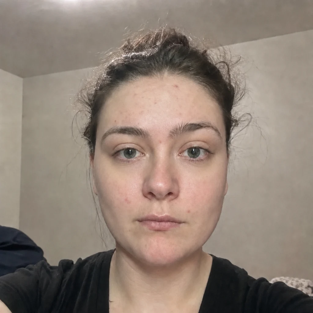
Before — generatedAfter — simulatedSee a orbital decompression on your own face
Run this simulationOrbital decompression risks, including double vision
- Double vision — new or worsened double vision after orbital decompression is a recognized risk, because moving the eye back changes how its muscles work. Its likelihood depends on which walls are decompressed and whether double vision was present before; it may settle or need eye muscle surgery.
- Numbness of the cheek, upper lip, gums or teeth, particularly after floor decompression; often improves over months.
- Sinus problems, including sinusitis, when the medial wall or floor is opened.
- Asymmetry or under- and over-correction of eye position.
- Bleeding and infection.
- Vision loss and cerebrospinal fluid leak — rare, but among the serious risks a surgeon will discuss.
Is orbital decompression worth it? For people with vision at risk, the question is usually settled by that risk. For proptosis in stable disease, it is a personal decision weighed with the surgeon, and patients commonly describe comfort and appearance as meaningful benefits — alongside the risks above.
Orbital decompression without surgery: medical treatment
There is no non-surgical way to enlarge the orbit, but thyroid eye disease itself is treated medically, and in some people that reduces swelling and proptosis enough that decompression is not needed:
- Steroids, often given intravenously during active disease, to reduce inflammation.
- Teprotumumab, an FDA-approved infused medication for thyroid eye disease that can reduce proptosis in some patients.
- Other immune-modulating treatments and orbital radiotherapy, used in selected cases.
- Supportive care — lubricating drops, sleeping with the head raised, protecting the eyes from wind and light, and in some mild cases selenium supplements.
- Stopping smoking, which is consistently associated with more severe thyroid eye disease.
- Thyroid level control, managed by an endocrinologist.
Filler and other cosmetic treatments do not move a protruding eye back. Some are used around the eyes to change how prominence reads, which is a different question from treating proptosis.
Orbital decompression cost, insurance and cosmetic cases
When orbital decompression is done for thyroid eye disease, it is generally treated as medical care and billed to health insurance, subject to the plan's rules, deductibles and prior authorization. Total cost varies widely with the number of walls, one or both eyes, the hospital and the surgical team, so there is no dependable single US price. Surgeons' offices and insurers can give an estimate for a specific plan.
Cosmetic orbital decompression
Some oculoplastic surgeons also perform orbital decompression for eyes that are naturally prominent without thyroid disease. It is the same operation, usually with a smaller amount of decompression, and carries the same categories of risk, including double vision and numbness. Cosmetic cases are not covered by insurance and are priced individually. Naturally prominent eyes are described on the protruding eyes page, which carries the same warning about prominence that has changed over time.
Orbital decompression simulation: what the render can and cannot show
The orbital decompression simulation settles protruding eyes back into the orbits so they sit more naturally in the face. For someone already on a treatment pathway, it can make a conversation about expected appearance more concrete.
It is the most limited render on this site. Proptosis is measured in millimeters with an instrument and planned from scans; a front-facing photo shows it poorly. The render does not show eyelid retraction, redness, double vision or the other procedures that often follow, and it cannot tell you why an eye is prominent. It is not a substitute for the assessment that thyroid eye disease requires. Lid surgery that often follows decompression is covered on the blepharoplasty page.
Questions to ask your oculoplastic surgeon about orbital decompression
- Is my thyroid eye disease active or stable, and is the aim to protect my vision, my eye's surface, or to reduce proptosis?
- Which walls and how much fat do you plan to decompress, how far do you expect the eye to move back, and what is my double vision risk with that plan?
- Where does this sit in the sequence of treatments I may need — medication, eye muscle surgery, eyelid surgery — and how many of these operations do you perform?
Alternatives to orbital decompression


How the orbital decompression simulation works
Your photo is analysed for the landmarks that matter to this procedure, and the render changes eye protrusion while leaving the rest of your face alone. That constraint is the hard part — a generic image model will happily redraw your whole face and hand you a stranger who looks better.
This is the procedure on this list where a render is least appropriate. It sits inside a medical pathway that starts with diagnosing why the eye is prominent, and no photograph starts that process.
It is an illustration of a plausible direction, not a prediction of your result and not a medical opinion. What it is genuinely good for is finding out whether you want the change at all — plenty of people render one, find the difference smaller than they imagined, and stop there.
Measure your face before a orbital decompression simulation
The free analysis tools measure symmetry, facial proportion and face shape, and tell you which measurements sit outside the reference range. Choosing a procedure from that is a much better starting point than choosing one from a photo you saved.



Orbital Decompression simulator — frequently asked questions
How painful is orbital decompression recovery?
It is usually described as pressure, aching and headache in the first days rather than severe pain, managed with prescribed medication. Swelling and bruising are the more noticeable part, easing over a few weeks.
What do thyroid eyes look like?
Common signs include eyes that look more prominent or staring, upper lids pulled higher so white shows above the iris, puffy lids, and red or irritated eyes. Many people with Graves' disease have mild or no eye changes.
How long does orbital decompression surgery take?
It depends on how many walls are decompressed and whether one or both eyes are treated, from roughly an hour to several hours. Your surgeon's plan gives the realistic estimate.
Can proptosis come back after decompression?
Once thyroid eye disease is inactive, the new eye position is usually stable. If the disease reactivates, swelling can return, which is one reason non-urgent surgery is typically planned for the stable phase.
Is orbital decompression covered by Medicare or insurance?
When it is performed for thyroid eye disease it is generally treated as medically necessary and can be covered, subject to the plan's requirements. Cosmetic orbital decompression is not covered.
Is the orbital decompression simulator free?
The face analysis is free and needs no account. A rendered simulation costs credits because it runs on a GPU — $4.99 buys five and does not require a subscription.
How accurate is an AI orbital decompression simulation?
It is an illustration of a plausible outcome, not a prediction of your result. Real outcomes depend on your anatomy, the oculoplastic surgeon and how you respond. Treat it as a conversation starter for a consultation, not a promise.
Do you keep my photo?
The free measurements never leave your browser. A simulation does send the photo to a rendering service, which is why it asks for explicit consent first, and you can delete the result from your dashboard at any time.
Can I show this to an oculoplastic surgeon?
Yes, and that is the most useful thing to do with it. Bringing a picture of the direction you want is far more precise than describing it in words.
Other eyes & brows simulations





This page is not medical advice and we are not clinicians. A simulation cannot tell you whether a procedure is appropriate for you, what it costs, or what the risks are. Those questions belong with a qualified oculoplastic surgeon.